- Published on
Detect COVID-19 with Deep Learning
- Authors
- Name
- Tan Ngoc Pham
- @ngctnnnn

Propose a rapidly testing method which has a high productivity in a short time, which is to use Deep Convolutional Neural Network to detect COVID-19 on Chest X-ray (CXR) images to cope with the present pandemic.
Table of contents
- Scientific base
- A deep learning based approach to the problem
- Experimental results and evaluation
- References
1. Scientific base
The most crucial thing that make CXR images from pneumonia or COVID-19 patients different from normal ones is the appearance of white spots, whether they are a lot of or a few, on particular positions along patients' lungs. Those white spots are recognized as the term of ground glass opacity or GGO in medical science. Ground glass opacity is the incompletely consolidated injury in patients' lungs. It has a higher density in comparison with surrounded parenchyma while still enables us to observe underlying structures, e.g. blood vessels or bronchial membranes.
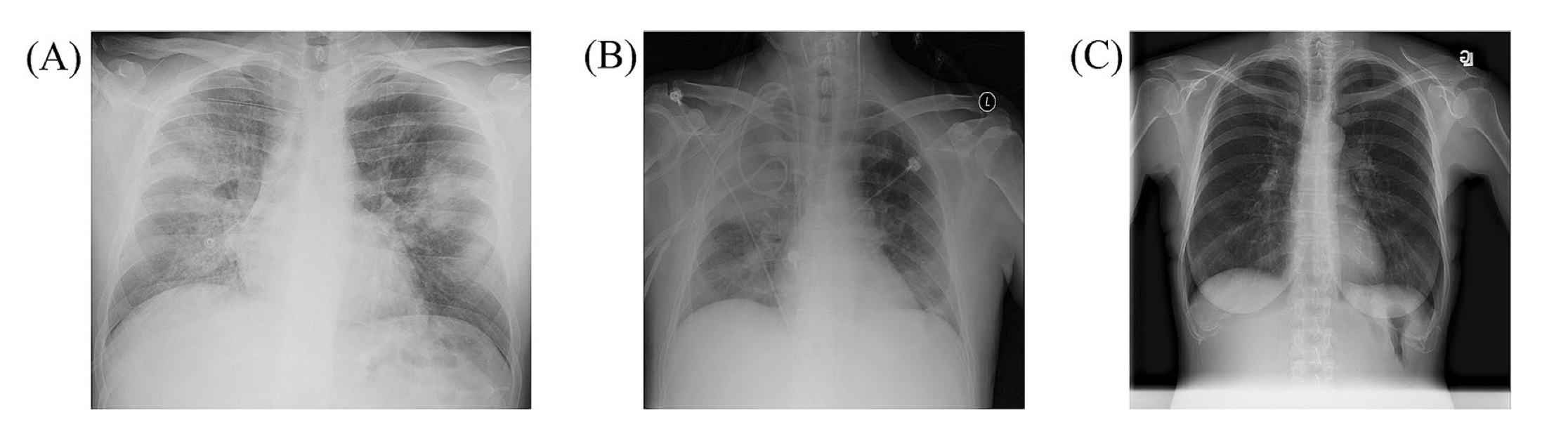
A specified doctor in the field of diagnostic imaging could tell that those GGO is the reason for those white spots in the chest radiograph. And a professional radiologist could use these features to differentiate COVID19 with pneumonia patients. Thus, we are capable of using a deep learning network to extract these features, then categorize to give out the appropriate diagnostic results for every cases.
2. A deep learning based approach to the problem
Throughout the research, we harness the use of 2 different approaches which are ResNet50 and VGG19 to solve this problem. In addtion, we use COVIDx dataset - which is a widely used dataset in recent research about COVID-19 nowadays.
VGG19 is a deep neural network architecture under-using residual design principals, it is also a compact architecture which has a low diversity of architectures. On the other hand, ResNet50 is a deep neural network harnessing residual design principles and it has a moderate diversity of architectures. This network brings many a high productivity in a large number of researching in classifying X-ray images.
2.1 COVIDx Dataset
COVIDx Datset is a dataset synthesized from 5 different sources. Additionally, this dataset also provides an image extension transfer tool: from .mri into .jpg. And the author moreover provide a code to support data pre-processing and getting rid of unnecessary part for synthesized data.
The dataset consists of more than 20.000 CXR images from different patients and divided into 2 sets which are training set and testing set. They are also separated into 3 classes which are pneumonia (train: 5963, test: 105), COVID-19 (train: 4649, test: 274) and the healthy (train: 8751, test: 100).
Our model will get an input of one CXR image and will give out an output as the probability of that image falling into each class which is pneumonia, COVID-19 and healthy, respectively.
2.2 Detailed implementation
Both deep learning neural network we proposed which are VGG19 and ResNet50 are all pre-trained on ImageNet. Afterwards, we proceed training process on COVIDx dataset with Adam as the optimization algorithm and the learning rate's strategy as reducing if the loss on validation set does not improve at all in a long period (patience).
Detailed implementation: ngctnnnn/Detect-COVID19.
After implementation, here is my demo for this project:
3. Experimental results and evaluation
| Disease | Precision | Recall | F1-score | Support |
|---|---|---|---|---|
| COVID-19 | 0.99 | 0.82 | 0.90 | 274 |
| Non-respiratory disease | 0.7 | 0.96 | 0.81 | 100 |
| Pneumonia | 0.8 | 0.86 | 0.83 | 105 |
| Disease | Precision | Recall | F1-score | Support |
|---|---|---|---|---|
| COVID-19 | 0.97 | 0.67 | 0.79 | 274 |
| Non-respiratory disease | 0.56 | 0.96 | 0.71 | 100 |
| Pneumonia | 0.74 | 0.85 | 0.79 | 105 |
| Disease | Precision | Recall | F1-score | Support |
|---|---|---|---|---|
| COVID-19 | 0.96 | 0.80 | 0.88 | 274 |
| Non-respiratory disease | 0.73 | 0.86 | 0.79 | 100 |
| Pneumonia | 0.71 | 0.90 | 0.79 | 105 |
| Architecture | Non-respiratory disease | Pneumonia | COVID-19 |
|---|---|---|---|
| VGG19 | 96% | 86% | 82% |
| ResNet50 (14 epochs) | 96% | 85% | 67% |
| ResNet50 (50 epochs) | 86% | 90% | 80% |
| Architecture | Non-respiratory disease | Pneumonia | COVID-19 |
|---|---|---|---|
| VGG19 | 70% | 80% | 99% |
| ResNet50 (14 epochs) | 56% | 74% | 97% |
| ResNet50 (50 epochs) | 73% | 71% | 96% |
| Architecture | Number of parameters (M) | Accuracy | Resolution |
|---|---|---|---|
| VGG19 | 29.76 trainable + 20.25 non-trainable | 86% | 480 x 480 |
| ResNet50 (14 epochs) | 25.93 trainable + 23.77 non-trainable | 77% | 224 x 224 |
| ResNet50 (50 epochs) | 25.93 trainable + 23.77 non-trainable | 84% | 224 x 224 |
4. References
[1] A. Chung. Actualmed covid-19 chest x-ray data initiative, 2020, URL: https://ncov.moh.gov.vn.
[2] J. P. Cohen et al. Covid-19 image data collection, 2020.
[3] J. Deng et al. Imagenet: A large-scale hierarchicalimage database. In2009 IEEE conference on computer vision and pattern recognition, pages 248–255.Ieee, 2009.
[4] K. He et al. Deep residual learning for image recognition, 2015.
[5] Ministry of Health of Vietnam. A page about acute respiratory tract infections covid-19, 2021. URL: https://ncov.moh.gov.vn.
[6] Radiological Society of North America. Covid-19 radiography database, 2019. URL: https://www.kaggle.com/tawsifurrahman/covid19-radiography-database.
[7] Radiological Society of North America. RSNA pneumonia detection challenge, 2019. URL: https://www.kaggle.com/c/rsna-pneumonia-detection-challenge/data.
[8] K. Simonyan and A. Zisserman. Very deep convolutional networks for large-scale image recognition, 2015.
[9] L. Wang, Z. Q. Lin, and A. Wong. Covid-net: a tailored deep convolutional neural network de-sign for detection of covid-19 cases from chest x-ray images.Scientific Reports, 10(1):19549,Nov 2020. ISSN 2045-2322. doi: 10.1038/s41598-020-76550-z. URL: https://doi.org/10.1038/s41598-020-76550-z.